The average cost of Tympanoplasty in India approximately ranges between INR 207875 to 313475 (USD 2500 to USD 3770)
Tympanoplasty (eardrum repair) refers to the surgical procedure that is performed to correct the ruptured eardrum or rebuild a perforated eardrum (tympanic membrane) or the middle ear’s tiny bones. Large defects caused due to severe infection or any accident require the surgery to restore the perforated eardrum to avoid further complications such as hearing loss. The procedure involves making a cut behind the ear to explore the middle ear. The graft of fascia or the cartilage is used to heal the hole in the eardrum. Either you bone or a synthetic bone is used to repair the bones of hearing. The surgeon closes the incision with stitches that are removed within 2 weeks of the operation. However, after four to six weeks after the operation, the patient needs to visit the doctor for a thorough hearing test.
With over 90 percent success rate of Tympanoplasty, India has all the latest medical advancements and technology, and skilled surgeons. Post-surgery care involves medications and some precautions.
India has the best facilities along with a skilled panel at very affordable rates. The expenses in India are 30% of that in most western countries. The cost of Eardrum surgery varies according to the severity of the condition and the method used for surgery. The cost of Tympanoplasty ranges from $2,700 to $4,100. The cost of the surgery in India is affordable with all the latest advancements, machines and technology, and the facilities provided by the hospitals are comparable to the best medical tourists’ destinations such as the US, UK, Mexico, Australia, and Canada.
| City | Minimum Cost | Maximum Cost |
|---|---|---|
| Gurgaon | USD 3040 | USD 3620 |
| Hyderabad | USD 3000 | USD 3540 |
| Panjim | USD 3010 | USD 3580 |
| Thane | USD 3290 | USD 3810 |
| Chennai | USD 3070 | USD 3710 |
| Pune | USD 3030 | USD 3820 |
| Kochi | USD 3070 | USD 3840 |
| Bengaluru | USD 3050 | USD 3540 |
| Ghaziabad | USD 3300 | USD 3760 |
| Greater Noida | USD 3280 | USD 3810 |
Treatment cost

The cost for Tympanoplasty ranges from USD 3300 - 3760 in Medanta - The Medicity
Medanta - The Medicity located in Gurugram, India is accredited by JCI, NABH. Also listed below are some of the most prominent infrastructural details:
DOCTORS IN 14 SPECIALITIES
FACILITIES & AMENITIES
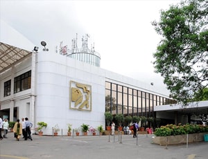
The cost for Tympanoplasty ranges from USD 3240 - 3560 in Apollo Hospitals
Apollo Hospitals located in Hyderabad, India is accredited by JCI, NABH. Also listed below are some of the most prominent infrastructural details:
DOCTORS IN 14 SPECIALITIES
FACILITIES & AMENITIES

The cost for Tympanoplasty ranges from USD 3220 - 3680 in Venkateshwar Hospital
Venkateshwar Hospital located in New Delhi, India is accredited by NABH. Also listed below are some of the most prominent infrastructural details:
DOCTORS IN 13 SPECIALITIES
FACILITIES & AMENITIES

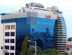
The cost for Tympanoplasty ranges from USD 3130 - 3840 in Star Hospitals
Star Hospitals located in Hyderabad, India is accredited by NABH, NABL. Also listed below are some of the most prominent infrastructural details:
DOCTORS IN 12 SPECIALITIES
FACILITIES & AMENITIES

The cost for Tympanoplasty ranges from USD 3260 - 3580 in Global Health City
The Hospital has world-class infrastructure with a capacity of over 1000 beds and much more-
DOCTORS IN 14 SPECIALITIES
FACILITIES & AMENITIES

The cost for Tympanoplasty ranges from USD 3270 - 3800 in Primus Super Speciality Hospital
Primus Super Speciality Hospital located in New Delhi, India is accredited by NABH. Also listed below are some of the most prominent infrastructural details:
DOCTORS IN 13 SPECIALITIES
FACILITIES & AMENITIES

The cost for Tympanoplasty ranges from USD 3150 - 3780 in Artemis Health Institute
Artemis Hospital is a 400 plus bed multi-speciality hospital, which aims at providing a depth of expertise in the spectrum of advanced medical and surgical interventions. Some of the features of the infrastructure include:
DOCTORS IN 15 SPECIALITIES
FACILITIES & AMENITIES

The cost for Tympanoplasty ranges from USD 3040 - 3570 in Sterling Wockhardt Hospital
Sterling Wockhardt Hospital located in Mumbai, India is accredited by NABH. Also listed below are some of the most prominent infrastructural details:
DOCTORS IN 14 SPECIALITIES
FACILITIES & AMENITIES

The cost for Tympanoplasty ranges from USD 3050 - 3700 in Nanavati Super Speciality Hospital
Nanavati Super Speciality Hospital located in Mumbai, India is accredited by NABH. Also listed below are some of the most prominent infrastructural details:
DOCTORS IN 14 SPECIALITIES
FACILITIES & AMENITIES
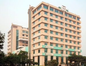
The cost for Tympanoplasty ranges from USD 3140 - 3640 in Max Super Speciality Hospital, Patparganj
Max Super Speciality Hospital, Patparganj located in New Delhi, India is accredited by JCI. Also listed below are some of the most prominent infrastructural details:
DOCTORS IN 14 SPECIALITIES
FACILITIES & AMENITIES

The cost for Tympanoplasty ranges from USD 3280 - 3750 in Apollo Hospitals Bannerghatta
Apollo Hospitals Bannerghatta located in Bengaluru, India is accredited by JCI, NABH. Also listed below are some of the most prominent infrastructural details:
DOCTORS IN 13 SPECIALITIES
FACILITIES & AMENITIES

The cost for Tympanoplasty ranges from USD 3100 - 3790 in Fortis Hospital
Fortis Hospital located in Bengaluru, India is accredited by ISO, NABH. Also listed below are some of the most prominent infrastructural details:
DOCTORS IN 12 SPECIALITIES
FACILITIES & AMENITIES
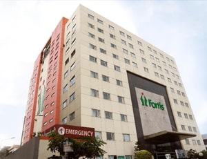
The cost for Tympanoplasty ranges from USD 3280 - 3680 in Fortis Hospital
Fortis Hospital located in Kolkata, India is accredited by ISO, NABH. Also listed below are some of the most prominent infrastructural details:
DOCTORS IN 12 SPECIALITIES
FACILITIES & AMENITIES

The cost for Tympanoplasty ranges from USD 3250 - 3780 in Sarvodaya Hospital and Research Centre
Sarvodaya Hospital and Research Centre located in Faridabad, India is accredited by NABH, NABL. Also listed below are some of the most prominent infrastructural details:
DOCTORS IN 14 SPECIALITIES
FACILITIES & AMENITIES
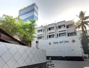
The cost for Tympanoplasty ranges from USD 3070 - 3560 in Ruby Hall Clinic
Ruby Hall Clinic located in Pune, India is accredited by NABH. Also listed below are some of the most prominent infrastructural details:
DOCTORS IN 13 SPECIALITIES
FACILITIES & AMENITIES
People from all age groups can be affected by ear disorders and hearing impairment problems. In the United States, a survey revealed that under the age of 65, over 60 percent of the population suffers from problems related to hearing loss. However, almost 25 percent of people aged above 65 experience a significant loss of hearing. Thankfully, different types of ENT surgeries are available to treat problems related to the ear.
Most often, people are diagnosed with problems related to the eardrum, or tympanum, and the infection in the cells of the mastoid bone. The tympanum is the thin membranous structure present between the outer and the middle ear. It vibrates when sound waves hit on it and this enables us to hear.
A repair surgery called tympanoplasty is required to treat a perforation or hole in the eardrum. The middle ear is sterile but due to rupture of the eardrum, an infection can also occur. It may also be required for repairing of tiny bones present behind the eardrum or ossicles in the mastoid bone. This repair is known as mastoidectomy.
Tympanoplasty and mastoidectomy are therefore conducted together in many cases. This procedure is known as tympano-mastoidectomy.
Several reasons may cause loss of hearing, including the following:
Birth defect
An ear infection that has grown very severe and left untreated for long
The injury suffered by the ear
Ear subjected to excessive levels of noise
Hearing loss as a result of age
Other reasons
Some of the common symptoms of a puncture in eardrum include the following:
Sharp pain in the ear that disappears abruptly
Excessive pressure, which will suddenly disappear with the rupture and pus formation in the ear
Loss of hearing
Dizziness
Type 1 tympanoplasty or myringoplasty is the surgery that ensures restoration of the eardrum that got perforated with drafting
Type II tympanoplasty addresses membrane perforations with erosion in the bone malleus. Grafting is done on the incus bone or in the remains of the malleus.
Type III tympanoplasty is meant for destruction of two ossicles and intact and mobile stapes bone. A graft is placed on the stapes and it provides protection for the total assembly.
Type IV tympanoplasty is useful for cases of ossicular destruction, which has all or part of the arch of stapes included. A graft is placed around the mobile stapes footplate
Type V tympanoplasty is useful when the footplate of the stapes bone is fixed.
For treating Cholesteatoma, tympanoplasty can be combined with stapedectomy and mastoidectomy and in many cases, a second operation is required to ensure the infection is totally eradicated.
The tympanoplasty surgery is performed with intravenous sedation and local anesthesia. An incision is made into the ear canal section and from the bony ear canal, the remaining eardrum is elevated and lifted forward. Under the operating microscope, the ear structures can be seen clearly. An incision behind the ear is made if the hole is very large or far forward. It ensures that the entire outer ear is forwarded, giving better access to the perforation.
The perforated remnant part is rotated forward after the hole is exposed and now the ossicles are inspected. Scar tissue and bands can surround the bones and they are removed with a laser or micro hooks. Now the ossicular chain is pressed to check its mobility and functionality. If it is found to be mobile, then the rest of the surgery aims at repairing the defect of the drum.
From the tragus, which is the cartilaginous lobe of skin in front of the ear, or from the back of the ear,
If the bones in the ear suffer from erosion, then ossicular reconstruction is advised. At times it can be determined before surgery but in other cases, erosion is visible only when the ear is completely opened under the microscope. The reconstruction can happen at the time of the eardrum construction. Bone erosion can happen at the tip of the incus or anvil. A discontinuity between the stapes and the incus has to be resolved.
A small piece of bone or cartilage can be inserted from some other part of the body of the patient if the gap between the two cones mentioned above is small. But if the gap is large, then the anvil bone is removed and remodeled to give a shape of a tooth with the help of the operating microscope. After reshaping the prosthesis, it is placed between the malleus and the stapes, and ossicular chain continuity is then re-established.
In some other ossicular construction, the malleus can get fixated by bony ingrowth or scar tissue to the ear’s lateral wall. The plastic-type or
Usually, a patient is discharged within two to three hours of the surgery. Along with a mild pain reliever, some antibiotics are also administered. After 10 days, the patient is again expected to visit so that packing can be removed and graft success can be checked.
Patients are advised to keep water away from the surgical site and avoid blowing of the nose. If the patient is suffering from cold and allergies, then decongestants are prescribed. Within 5 to 6 days, the patients can resume a normal life. After 3 weeks of the surgery, the packing is removed under the operating microscope and at this point, the grafting success can be completely determined.
Care must be taken by the patient to soak the ear canal with antibiotics to keep infection at bay. Shearing forces of excessive tension should not be felt by the graft. The surgeon will advise the patient to avoid activities that alter the tympanic pressure, including using a straw to drink or blowing of the nose. Finally, a hearing test is performed after 4 to 6 weeks of the surgery.
Ask your healthcare adviser for the best multiple options and choose the one that meets your expectations
The average cost of Tympanoplasty in India starts from USD 2300 In India, Tympanoplasty is conducted across many multispecialty hospitals.
The cost of Tympanoplasty in India may differ from one medical facility to the other. The Tympanoplasty package cost usually includes all the expenses related to pre and post surgery expenses of the patient. The Tympanoplasty cost in India includes the cost of anesthesia, medicines, hospitalization and the surgeon's fee. A prolonged hospital stay due to delayed recovery, new diagnosis and complications after surgery may increase the cost of Tympanoplasty in India.
There are several best hospitals for Tympanoplasty in India. For quick reference, the following are some of the leading hospitals for Tympanoplasty in India:
The recovery of the patient many vary, depending on several factors. However, on an average, patient is supposed to stay for about 10 days in the country after discharge. This time frame is important to ensure that the surgery was successful and the patient is fit to fly back.
Apart from the Tympanoplasty cost, there are a few other daily charges that the patient may have to pay. These are the charges for daily meals and accommodation outside the hospital. The per day cost in this case may start from USD 50 per person.
There are many cities that offer Tympanoplasty in India, including the following:
Patients can also avail to attend a video teleconsultation with the Tympanoplasty surgeon in India. the following are some of the top doctors offering Tympanoplasty in India:
| Doctor | Cost | Schedule Your Appointment |
|---|---|---|
| Dr. Dhirendra Singh Khushwah | USD 13 | Schedule Now |
The patient is supposed to stay at the hospital for about 2 days after Tympanoplasty for monitoring and care. This phase is important to ensure that the patient is recovering well and is clinically stable. During this time, several tests are performed before the patient is deemed suitable for discharge.
The average rating for Tympanoplasty hospitals in India is 3.9. This rating is automatically calculated on the basis of several parameters such as the infrastructure of the hospital, quality of services, nursing support and other services.
There are more than 51 hospitals that offer Tympanoplasty in India. These hospitals have proper infrastructure for the treatment of patients who require kidney transplant. These hospitals comply with all the rules and regulations as dictated by the regulatory bodies and medical association in India
Some of the top doctors for Tympanoplasty in India are: