A craniotomy is a major brain surgery in which a bone flap from the skull is temporarily removed to have an access to the brain. This procedure is mainly performed in patients who suffer from traumatic brain injuries or brain lesions. It is a highly critical procedure and a patient may take months to recover from it. This surgery is also conducted prior to the placement of deep brain stimulators, which is often recommended to patients suffering from epilepsy and Parkinson’s disease. This procedure has a wide range of other applications as well such as brain imaging and electrical stimulation.
A craniotomy is a highly sensitive procedure that is performed in some of the top neurosurgery hospitals in U.A.E. Because of the sensitivity of the procedure and the risks involved, this surgery should only be conducted by highly experienced and skilled surgeons. The top neurosurgeons in U.A.E are educated from some of the most prestigious medical universities in the world. They hold several years of experience in conducting all types of brain surgeries and have so far treated thousands of patients from around the world.
Treatment cost

Types of Craniotomy in Zulekha Hospital Sharjah and its associated cost
| Treatment Option | Approximate Cost Range (USD) | Approximate Cost Range (AED) |
|---|---|---|
| Overall Craniotomy Surgery | 15091 - 28645 | 56626 - 102381 |
| Extended Bifrontal Craniotomy | 11093 - 27286 | 39272 - 101823 |
| Supra-Orbital Craniotomy | 11828 - 24608 | 42422 - 89575 |
| Retro Sigmoid Craniotomy | 13546 - 27010 | 48994 - 97772 |
| Orbitozygomatic Craniotomy | 13439 - 26931 | 50769 - 98755 |
| Translabyrinthine Craniotomy | 13784 - 28077 | 50166 - 103627 |
| Pterional Craniotomy | 12280 - 26860 | 45411 - 97895 |
| Suboccipital Craniotomy | 12546 - 25020 | 44467 - 91563 |

Types of Craniotomy in Zulekha Hospital Dubai and its associated cost
| Treatment Option | Approximate Cost Range (USD) | Approximate Cost Range (AED) |
|---|---|---|
| Overall Craniotomy Surgery | 15425 - 28706 | 56346 - 102603 |
| Extended Bifrontal Craniotomy | 10721 - 27813 | 39598 - 98383 |
| Supra-Orbital Craniotomy | 11542 - 24304 | 41750 - 91089 |
| Retro Sigmoid Craniotomy | 13721 - 27308 | 50486 - 101049 |
| Orbitozygomatic Craniotomy | 13459 - 27790 | 50799 - 98613 |
| Translabyrinthine Craniotomy | 13381 - 28610 | 49841 - 105383 |
| Pterional Craniotomy | 12423 - 26342 | 44412 - 100853 |
| Suboccipital Craniotomy | 12471 - 25001 | 45638 - 95171 |
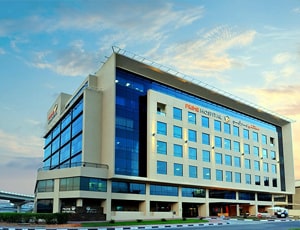
Types of Craniotomy in Prime Hospital and its associated cost
| Treatment Option | Approximate Cost Range (USD) | Approximate Cost Range (AED) |
|---|---|---|
| Overall Craniotomy Surgery | 15031 - 27737 | 56263 - 106132 |
| Extended Bifrontal Craniotomy | 10687 - 27232 | 40604 - 100516 |
| Supra-Orbital Craniotomy | 11543 - 23989 | 42629 - 89043 |
| Retro Sigmoid Craniotomy | 13544 - 26654 | 50437 - 100508 |
| Orbitozygomatic Craniotomy | 13884 - 26799 | 50607 - 101327 |
| Translabyrinthine Craniotomy | 13820 - 28312 | 48929 - 104010 |
| Pterional Craniotomy | 12161 - 27423 | 44534 - 98491 |
| Suboccipital Craniotomy | 12333 - 25804 | 46174 - 92732 |

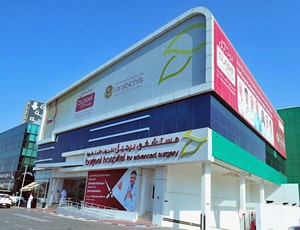
Types of Craniotomy in Burjeel Hospital for Advanced Surgery Dubai and its associated cost
| Treatment Option | Approximate Cost Range (USD) | Approximate Cost Range (AED) |
|---|---|---|
| Overall Craniotomy Surgery | 15461 - 28383 | 56949 - 106218 |
| Extended Bifrontal Craniotomy | 10957 - 27652 | 39659 - 99991 |
| Supra-Orbital Craniotomy | 11609 - 24732 | 42073 - 90183 |
| Retro Sigmoid Craniotomy | 13426 - 26737 | 50429 - 99821 |
| Orbitozygomatic Craniotomy | 13346 - 26898 | 49526 - 98549 |
| Translabyrinthine Craniotomy | 13365 - 28822 | 50132 - 106363 |
| Pterional Craniotomy | 12281 - 26938 | 45422 - 97505 |
| Suboccipital Craniotomy | 12481 - 25382 | 45639 - 92433 |

Types of Craniotomy in Iranian Hospital and its associated cost
| Treatment Option | Approximate Cost Range (USD) | Approximate Cost Range (AED) |
|---|---|---|
| Overall Craniotomy Surgery | 15100 - 28482 | 54874 - 105977 |
| Extended Bifrontal Craniotomy | 10852 - 27702 | 39998 - 101991 |
| Supra-Orbital Craniotomy | 11684 - 24128 | 42364 - 91127 |
| Retro Sigmoid Craniotomy | 13846 - 27376 | 50050 - 98361 |
| Orbitozygomatic Craniotomy | 13702 - 26785 | 49282 - 100573 |
| Translabyrinthine Craniotomy | 13848 - 28219 | 49625 - 105196 |
| Pterional Craniotomy | 12557 - 27315 | 46424 - 98430 |
| Suboccipital Craniotomy | 12123 - 25046 | 45191 - 93352 |
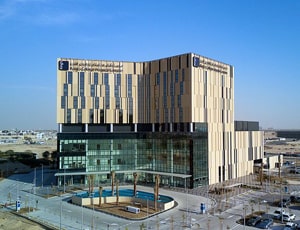
Types of Craniotomy in Kings College Hospital Dubai and its associated cost
| Treatment Option | Approximate Cost Range (USD) | Approximate Cost Range (AED) |
|---|---|---|
| Overall Craniotomy Surgery | 15421 - 28103 | 56795 - 104188 |
| Extended Bifrontal Craniotomy | 11125 - 27022 | 40030 - 99335 |
| Supra-Orbital Craniotomy | 11489 - 24607 | 43018 - 89123 |
| Retro Sigmoid Craniotomy | 13332 - 26623 | 50881 - 101276 |
| Orbitozygomatic Craniotomy | 13637 - 27796 | 49480 - 101021 |
| Translabyrinthine Craniotomy | 13592 - 29043 | 49283 - 105768 |
| Pterional Craniotomy | 12455 - 26980 | 45106 - 96498 |
| Suboccipital Craniotomy | 12398 - 25647 | 45070 - 93989 |
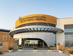
The Neuro Spinal Hospital is located in Dubai Science Park in Dubai. It is a pioneer in bringing the latest Technology, Medicine, and Education to heal and serve the community. It has been approved and accredited by the Dubai Health Authority and has the Government of Dubai and Dubai Health Experience as its partners.
It has a 114-bed capacity, green spaces for the rehabilitation of the patients, and smart patient rooms, and is four times that of its former premises in terms of capacity. It is also the first hospital to have surgical robots in UAE. The Continous Medical Education (CME) program was introduced in 2007 by the hospital that has become a mandatory requirement for all healthcare professionals. Practitioners need to provide that they have attended enough CME hours to be promoted to a higher level or renew their licenses.
The hospital consists of multinational staff who are dedicated to working together for solving problems with the patients and their families, to provide the highest level of support and care through advanced and progressive technologies, and an evidence approach to medicine, all delivered in a collaborative and compassionate environment. The team believes in safety, quality, dignity, engagement, and collaboration in providing premium healthcare to the patient. It consists of specialized units such as spinal and back pain units, joint replacement centers, sports medicine, orthopedics, oncology, neurology, physiotherapy, etc.
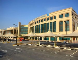
Types of Craniotomy in NMC Royal Hospital, Khalifa City and its associated cost
| Treatment Option | Approximate Cost Range (USD) | Approximate Cost Range (AED) |
|---|---|---|
| Overall Craniotomy Surgery | 15428 - 28424 | 55378 - 105496 |
| Extended Bifrontal Craniotomy | 10847 - 27624 | 39439 - 98433 |
| Supra-Orbital Craniotomy | 11663 - 24033 | 43199 - 89821 |
| Retro Sigmoid Craniotomy | 13375 - 27396 | 49460 - 99232 |
| Orbitozygomatic Craniotomy | 13865 - 27374 | 50810 - 98898 |
| Translabyrinthine Craniotomy | 13875 - 28950 | 49482 - 104820 |
| Pterional Craniotomy | 12105 - 26869 | 44893 - 97920 |
| Suboccipital Craniotomy | 12496 - 25572 | 45537 - 94282 |
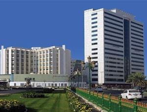
Types of Craniotomy in NMC Royal Hospital Sharjah and its associated cost
| Treatment Option | Approximate Cost Range (USD) | Approximate Cost Range (AED) |
|---|---|---|
| Overall Craniotomy Surgery | 15024 - 27977 | 56795 - 102496 |
| Extended Bifrontal Craniotomy | 10691 - 27305 | 40172 - 98914 |
| Supra-Orbital Craniotomy | 11832 - 24080 | 42401 - 89524 |
| Retro Sigmoid Craniotomy | 13667 - 26709 | 49797 - 101441 |
| Orbitozygomatic Craniotomy | 13567 - 26756 | 49270 - 101334 |
| Translabyrinthine Craniotomy | 13750 - 28131 | 48891 - 102408 |
| Pterional Craniotomy | 12500 - 26804 | 45165 - 100746 |
| Suboccipital Craniotomy | 12155 - 25015 | 45149 - 94277 |
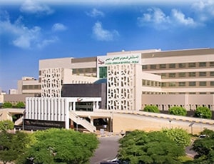
Types of Craniotomy in Saudi German Hospital and its associated cost
| Treatment Option | Approximate Cost Range (USD) | Approximate Cost Range (AED) |
|---|---|---|
| Overall Craniotomy Surgery | 15140 - 28370 | 55626 - 105458 |
| Extended Bifrontal Craniotomy | 10860 - 27792 | 40188 - 99048 |
| Supra-Orbital Craniotomy | 11690 - 24558 | 43009 - 88814 |
| Retro Sigmoid Craniotomy | 13826 - 27639 | 50583 - 101269 |
| Orbitozygomatic Craniotomy | 13353 - 27586 | 50636 - 101439 |
| Translabyrinthine Craniotomy | 13735 - 28023 | 50599 - 104978 |
| Pterional Craniotomy | 12145 - 27416 | 44512 - 96838 |
| Suboccipital Craniotomy | 12240 - 25861 | 46333 - 92617 |

Types of Craniotomy in LLH Hospital, Abu Dhabi and its associated cost
| Treatment Option | Approximate Cost Range (USD) | Approximate Cost Range (AED) |
|---|---|---|
| Overall Craniotomy Surgery | 14917 - 28597 | 54882 - 104044 |
| Extended Bifrontal Craniotomy | 10848 - 26927 | 40401 - 100747 |
| Supra-Orbital Craniotomy | 11347 - 24377 | 42172 - 91875 |
| Retro Sigmoid Craniotomy | 13329 - 26943 | 50830 - 101421 |
| Orbitozygomatic Craniotomy | 13593 - 27195 | 50066 - 101282 |
| Translabyrinthine Craniotomy | 13845 - 28918 | 49526 - 102723 |
| Pterional Craniotomy | 12541 - 27390 | 45011 - 100609 |
| Suboccipital Craniotomy | 12637 - 25119 | 45443 - 91351 |
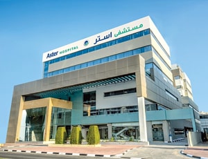
Types of Craniotomy in Aster DM Healthcare and its associated cost
| Treatment Option | Approximate Cost Range (USD) | Approximate Cost Range (AED) |
|---|---|---|
| Overall Craniotomy Surgery | 15127 - 27799 | 56170 - 103286 |
| Extended Bifrontal Craniotomy | 11144 - 27062 | 40233 - 100718 |
| Supra-Orbital Craniotomy | 11387 - 24659 | 42602 - 90874 |
| Retro Sigmoid Craniotomy | 13847 - 27369 | 50037 - 99188 |
| Orbitozygomatic Craniotomy | 13406 - 27061 | 49269 - 98464 |
| Translabyrinthine Craniotomy | 13780 - 28450 | 50000 - 105849 |
| Pterional Craniotomy | 12489 - 26519 | 45866 - 96849 |
| Suboccipital Craniotomy | 12384 - 25096 | 46211 - 91886 |
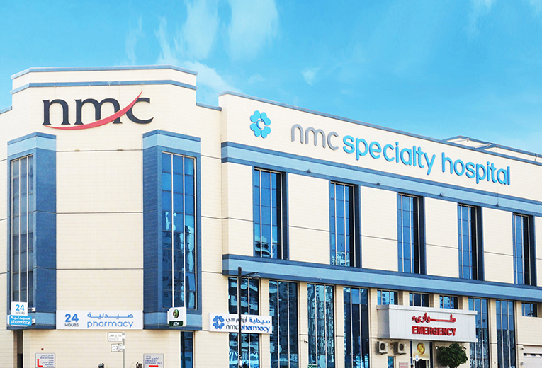
Types of Craniotomy in NMC Specialty Hospital, Al Nahda and its associated cost
| Treatment Option | Approximate Cost Range (USD) | Approximate Cost Range (AED) |
|---|---|---|
| Overall Craniotomy Surgery | 15349 - 28423 | 55640 - 101982 |
| Extended Bifrontal Craniotomy | 11071 - 26852 | 40621 - 100963 |
| Supra-Orbital Craniotomy | 11694 - 24920 | 42160 - 88845 |
| Retro Sigmoid Craniotomy | 13464 - 27784 | 50003 - 98379 |
| Orbitozygomatic Craniotomy | 13461 - 27567 | 50070 - 98983 |
| Translabyrinthine Craniotomy | 13313 - 28821 | 50112 - 104246 |
| Pterional Craniotomy | 12398 - 26580 | 45226 - 98647 |
| Suboccipital Craniotomy | 12199 - 24935 | 46352 - 93091 |
Types of Craniotomy in Lifecare Hospital, Musaffah and its associated cost
| Treatment Option | Approximate Cost Range (USD) | Approximate Cost Range (AED) |
|---|---|---|
| Overall Craniotomy Surgery | 15018 - 28427 | 55898 - 103058 |
| Extended Bifrontal Craniotomy | 10956 - 27349 | 39569 - 97873 |
| Supra-Orbital Craniotomy | 11841 - 24579 | 42401 - 91667 |
| Retro Sigmoid Craniotomy | 13364 - 27779 | 48988 - 100289 |
| Orbitozygomatic Craniotomy | 13877 - 26787 | 49091 - 99927 |
| Translabyrinthine Craniotomy | 13878 - 29058 | 50513 - 104469 |
| Pterional Craniotomy | 12218 - 26517 | 45342 - 99939 |
| Suboccipital Craniotomy | 12316 - 25888 | 44707 - 93404 |

Types of Craniotomy in Thumbay University Hospital, Ajman and its associated cost
| Treatment Option | Approximate Cost Range (USD) | Approximate Cost Range (AED) |
|---|---|---|
| Overall Craniotomy Surgery | 15303 - 28082 | 55401 - 105720 |
| Extended Bifrontal Craniotomy | 10773 - 27630 | 39378 - 101593 |
| Supra-Orbital Craniotomy | 11339 - 24171 | 43055 - 90638 |
| Retro Sigmoid Craniotomy | 13709 - 27825 | 49544 - 99889 |
| Orbitozygomatic Craniotomy | 13602 - 27617 | 49565 - 98260 |
| Translabyrinthine Craniotomy | 13527 - 28705 | 49858 - 103614 |
| Pterional Craniotomy | 12132 - 26430 | 46268 - 98568 |
| Suboccipital Craniotomy | 12350 - 25432 | 45223 - 95148 |
Craniotomy surgery is one of the most common types of brain surgery conducted to treat a brain tumor. It mainly aims at removing a lesion, tumor, or a blood clot in the brain by opening a flap above the brain to access the targeted area. This flap is removed on a temporary basis and again put in place when the surgery is done. Around 90 percent of the cases of brain tumors are diagnosed in adults aged between 55 and 65. Among children, a brain tumor is diagnosed within an age range of 3 to 12 years.
Craniotomy procedures are conducted with the help of magnetic resonance imaging (MRI) scans to reach the location precisely in the brain that requires treatment. A three-dimensional image for the same is achieved of the brain in conjunction with localizing frames and computers to view a tumor properly. A clear distinction is made between abnormal or tumor tissue and normal healthy tissue and to access the exact location of the abnormal tissue.
In a minimally invasive craniotomy procedure, a burr hole or a keyhole may be created to access the brain to fulfill the following purposes:
When there are complex craniotomies involved, the procedure may be referred to as a skull base surgery. In this kind of surgery, a small portion of the skull is removed from the bottom of the brain. This is the region where delicate arteries, veins, and cranial nerves exit the skull. Complicated planning is done to plan such craniotomies and understand the location of the lesions. This type of approach is usually employed for:
Primary brain tumors are much less common than secondary brain tumors. Primary ones are found to originate very close to the brain itself or in the tissues very close to it, such as the covering membranes of the brain, including the meninges, cranial nerves, pineal, or pituitary gland. It begins with normal cells, which at a later period undergoes some mutational errors in their DNA. The mutation triggers cells to grow and divide at a very high rate while healthy cells keep dying around it. This results in a mass of abnormal cells which gives rise to a tumor. Unlike primary tumors, the secondary tumors begin as cancer elsewhere and spread to the brain.
No matter what the goal of the surgery is, it is best to ensure that the incision is made to address the intracranial lesion keeping some principles in mind. A wide variety of intracranial processes can be done via a craniotomy with a different variety of incisions. Some of these variations include frontal craniotomy, pterional craniotomy, temporal craniotomy, decompression craniectomy, and suboccipital craniotomy.
Ask your healthcare adviser for the best multiple options and choose the one that meets your expectations
The cost of Craniotomy in the United Arab Emirates may differ from one medical facility to the other. The top hospitals for Craniotomy in the United Arab Emirates covers all the expenses related to the pre-surgery investigations of the candidate. The Craniotomy cost in the United Arab Emirates includes the cost of anesthesia, medicines, hospitalization and the surgeon's fee. Stay outside the package duration, post-operative complications and diagnosis of a new condition may further increase the Craniotomy cost in the United Arab Emirates.
There are many hospitals across the country that offer Craniotomy to international patients. Some of the most renowned hospitals for Craniotomy in the United Arab Emirates include the following:
After Craniotomy in the United Arab Emirates, the patient is supposed to stay in guest house for another 28 days. This time frame is important to ensure that the surgery was successful and the patient is fit to fly back.
There are certain expenses additional to the Craniotomy cost that the patient may have to pay for. These are the chanrges for daily meals and hotel stay outside the hospital. These charges starts from USD 50 per person.
The following are some of the best cities for Craniotomy in the United Arab Emirates:
Patients who are interested in availing telemedicine consultation before they travel for Craniotomy in the United Arab Emirates can opt for the same. There are many Craniotomy surgeons who offer video telemedicine consultation, including the following:
| Doctor | Cost | Schedule Your Appointment |
|---|---|---|
| Dr. Mehandi Hassan Ansari | USD 173 | Schedule Now |
| Dr. Ajit Kumar | USD 173 | Schedule Now |
| Dr. Shankar Ayyappan Kutty | USD 173 | Schedule Now |
| Dr. Rahul Amunje Mally | USD 173 | Schedule Now |
| Dr. Arif Khan | USD 173 | Schedule Now |
The patient has to spend about 5 days in the hospital after Craniotomy for proper recovery and to get clearance for discharge. During the recovery, the patient is carefully monitored and control tests are performed to see that everything is okay. If required, physiotherapy sessions are also planned during recovery in hospital.
The average rating for Craniotomy hospitals in the United Arab Emirates is 4.5. Several parameters such as hospital infrastructure, pricing policy, quality of services, politeness of staff etc. contribute to the rating.
There are more than 19 hospitals that offer Craniotomy in the United Arab Emirates. These clinics have propoer infrastructure as well as offer good quality of services when it comes to Craniotomy Apart from good services, the hospitals are known to follow all standard and legal guidelines as dictated by the local medical affairs body or organization.
Some of the most sought after medical specialists for Craniotomy in the United Arab Emirates are: