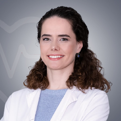
10 Years of experience
Speaks: English
A neurosurgeon is a medical specialist who diagnoses and treats conditions affecting the nervous system, including the brain, peripheral nerves, spinal cord, and spine. Neurosurgeons offer non-operative as well as surgical treatment to patients. Here is a list of some of the conditions Dr. Semra Isik treats:
Some of the signs and symptoms that neurological disorders can produce are listed below. These symptoms should be discussed with a neurosurgeon for further medical investigation.
The above symptoms appear mainly due to conditions that can adversely affect the nervous system. The symptoms can vary from person to person because the nervous system regulates different body functions. Symptoms can include all forms of pain.
If you wish to see Dr Semra Isik in his clinic/hospital, you can visit him between 11 am and 5 pm (Monday to Saturday). The doctor doesn’t work on Sunday. Call the doctor or his attendant to confirm his/her availability because the doctor may not be available due to some personal reasons or any emergencies.
Some of the popular procedures that Dr Semra Isik perform are given below::
One of the eminent neurosurgeons in the world, Dr. Semra Isik has specialized in the surgical treatment of diseases of the nervous system. The doctor works with a team of highly experienced physicians and can handle even the most complex with quite an ease. The neurosurgeon follows all medical protocols to ensure patient safety.

Share Your Experience about Dr. Semra Isik

Neurosurgeons, also known as brain surgeons, are doctors who specialize in the surgical treatment of conditions that affect the nervous system, brain, and spine. Neurosurgeons first have the training which makes them eligible to practise as a doctor. After this, they complete specialist training in neurosurgery. Neurosurgeons mostly see patients in their own clinics and public and private hospitals. Sometimes, they have to work with other specialists and medical experts to seek their opinion on diagnosis and surgery techniques. They also evaluate the diagnostic tests to know the exact underlying conditions and accordingly proceed with the treatment.
Diagnosis tests act as an important tool to find out the condition(s) a patient is suffering from. So, a neurosurgeon will ask you to get a few tests done so that they get to know the cause of the symptoms which further helps in finding the condition the patient is suffering from. Based on the diagnosis, the doctor can start appropriate treatment. A neurological examination is the assessment of motor responses and sensory neuron and includes the following:
Below are some tests that a neurosurgeon may recommend for the diagnosis of the conditions of the nervous system.:
Here are some of the common signs that you must seek the medical assistance of a neurosurgeon:
Neurosurgeons help in the diagnosis and treatment of the conditions of the nervous system. They are mostly involved in complicated surgery of the brain. They offer surgical treatment for the conditions affecting any part of the body, caused mainly due to nerve issues.