Vitreous Hemorrhage Treatment in Turkey
- Top Accredited Hospitals
- Global Medical Standards
- Cutting-Edge Technology
- Proven Treatment Options
- High Success Rate
- Trusted by Millions

What is a vitreous haemorrhage?
A vitreous haemorrhage is a condition in which blood leaks into the vitreous, the clear, gel-like fluid inside the eye. It can happen when blood vessels in the retina or nearby tissues bleed, often due to diabetic retinopathy, retinal tears or detachment, eye trauma, or other retinal disorders. Blood can block light from reaching the retina, causing sudden vision changes. Prompt assessment is needed to determine the appropriate treatment.
What is the Importance of Timely Treatment?
Timely treatment matters because a vitreous haemorrhage may signal an underlying retinal rupture, retinal detachment, or other serious eye problem; prompt evaluation and treatment are crucial. Early evaluation can reduce the risk of irreversible vision loss and help determine the cause. Treatment options depend on the cause and severity and may include vitrectomy, laser therapy, injections, or observation.
What are the common symptoms of a vitreous haemorrhage?
- Sudden blurred or reduced vision
- New or numerous small dots in vision.
- Dark spots or shadows
- Cobweb-like structure.
- Flashes of light.
- Vision may become very hazy or significantly reduced.
- curtain-like shadow
Causes and Risk Factors of Vitreous Haemorrhage
Causes
- Retinal tears or detachment: A retinal tear may damage blood vessels and cause bleeding. Retinal detachment may occur alongside vitreous haemorrhage.
- Posterior vitreous detachment: Age-related separation of the vitreous from the retina can sometimes cause bleeding, particularly if a retinal tear develops.
- Retinal vein occlusion: A blocked retinal vein may lead to abnormal blood vessel growth and bleeding.
- Eye trauma: An injury to the eye may damage retinal or other intraocular blood vessels.
- Retinal vascular inflammation or other vascular disorders: Certain conditions can damage blood vessels and cause bleeding.
- Retinal tumours or other uncommon eye disorders: Some may cause bleeding within the eye.
Risk Factors
- Long-standing diabetes, particularly with diabetic retinopathy.
- High blood pressure, especially when associated with retinal vascular disease
- Retinal tears or detachment
- Age-related changes in the vitreous fluid
- Blood-clotting disorders, or blood-thinning medicines
- Previous retinal disease
Latest Research and Technologies in the Treatment of Vitreous Haemorrhage in turkey
- Turkish research has evaluated this technique's outcomes in patients with advanced diabetic eye disease. Anti-VEGF injections may also be used in selected cases of diabetic retinopathy to reduce abnormal blood vessel activity and help manage bleeding around surgery. Optical coherence tomography (OCT) and B-scan ultrasonography support diagnosis and treatment planning, particularly when retinal structures need detailed assessment or bleeding obscures the retina. Treatment choice depends on the cause of the haemorrhage, retinal findings, and the patient's visual needs
Treatment options for Vitreous Hemorrhage
Vitrectomy : is the procedure in which the surgeon removes the damaged vitreous gel from inside the eye. During the procedure, the surgeon removes the blood-filled vitreous gel and replaces it with a clear fluid or another suitable material, such as saline, silicone oil, or a gas bubble.
Cost Start From 3500Explore Options
Laser photocoagulation: It uses focused laser light to close leaking or abnormal blood vessels in the retina or to fix a retinal tear. In vitreous haemorrhage, it mainly treats the cause of the bleeding and helps prevent further bleeding. This procedure is usually done as an outpatient treatment after numbing the eye.
- Clinical assessment:
- Medical examination: The physician may request a comprehensive medical history and a summary of the patient's signs and symptoms.
- Slit lamp examination: A slit lamp examination or an ophthalmoscope is used to check for any signs of bleeding.
- Dilated retinal examination: The pupil is widened with eye drops to allow the ophthalmologist to examine the retina for bleeding, retinal tears, or other abnormalities, where visibility permits.
- Imaging test:
- B-scan ultrasonography: enables examination of the retina when dense bleeding obstructs a clear view and can identify retinal tears or detachments.
- Optical Coherence Tomography (OCT): Provides detailed images of the retina when it can be visualised.
- Imaging scans: Computed tomography and magnetic resonance imaging can assess bleeding inside your eye.
false
- No medication directly treats vitreous hemorrhage. Medication therapy mainly targets the underlying cause; for example, anti-VEGF injections help reduce abnormal blood vessel growth and bleeding. Corticosteroids may be used in selected retinal conditions to reduce inflammation or retinal swelling. The choice depends on the diagnosis and should be determined by an ophthalmologist.
Instantly Connect with our Specialists


Hospitals for Vitreous Hemorrhage in Turkey

Can Hospital
Izmir, Turkey
One of Turkey's leading hospitals, Can Hospital in Izmir, was established in 2017 and has a 95-bed capacity. It has been accredited by the International Organization for Standardization (ISO), the International Health Tourism Authorization Certificate, the Turkish Republic Ministry Of Health Accreditation, the Turkish Republic Service Exporters' Association, the Association Of Turkish Travel Agencies, and the Turkish Republic Ministry Of Health Portal.
The hospital offers excellent, cutting-edge facilities for the patients' comfortable stay. They offer various medical services, including eye surgery, dentistry, hair transplantation, cosmetic and reconstructive surgery, and surgeries for metabolic and obesity disorders. The hospital upholds the highest standards for hygiene, has exceptional patient care, and employs specialist technology.
The hospital's doctors, nurses, and medical technicians are highly skilled and available around the clock for the patients. The hospital's physicians and surgeons are dedicated to offering the most outstanding medical care at reasonable costs. The hospital offers language services such as Turkish, English, German, French, and Spanish.
To offer a top-notch patient experience throughout their stay in the hospital, it has 350 workers working diligently. The hospital has specialized divisions such as Dentistry, Cosmetic and Plastic Surgery, Hair Clinic, Ophthalmology, Orthopedics, ENT treatment, and Obesity or Bariatric Surgery.
Our Services to better your experience
Opinion & Option
We submit the most accurate opinion and options from one or more countries for your review
Consult Privately
Consult with a certified specialist privately on our telemedicine platform even before you decide to travel
Logistics
We handle flights, visas, transfers, and accommodation—so you can focus on your health.
Recovery
Our In-house rehabilitation service packages to better your recovery and treatment outcome
Why Choose Turkey for Vitreous Haemorrhage Treatment?
- Accredited hospital: Turkey has advanced, highly specialised top-tier JCI- and NABH-accredited eye hospitals that use advanced optical coherence tomography (OCT) and high-success surgical techniques.
- Specialised surgeons: Turkey's vitreoretinal surgeons routinely handle complex cases using international protocols.
- Modern diagnostic procedures: Patients can access advanced ophthalmic procedures, and specialised centres offer techniques to support diagnosis and treatment planning.
- Coordinated follow-up: Patients can ask whether the same ophthalmology service provides diagnosis, treatment, and postoperative monitoring.
- International patient support: Some hospitals offer appointment coordination and assistance with travel arrangements. Availability varies between providers.
Frequently Asked Questions

Reviewer
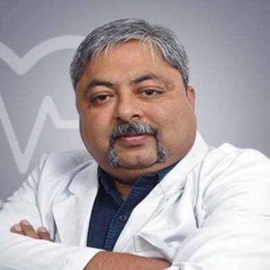
Dr. Vidit Tripathi
ENT Surgeon
24 Years of Experience
One of the finest ENT Surgeon in New Delhi, India, Dr. Vidit Tripathi, has worked with several world class multidisciplinary hospitals over the years. Dr. Vidit Tripathi has over 24+ years of experience in his field. The specialist treats and manages a wide range of conditions such as Goiter, Radiation Necrosis in Neck, Neck Injury, Perforates Eardrum View More
Last Reviewed - January 2026