Ureterolithiasis (Ureteral Stones) Treatment in Malaysia
- Top Accredited Hospitals
- Global Medical Standards
- Cutting-Edge Technology
- Proven Treatment Options
- High Success Rate
- Trusted by Millions

What is Ureterolithiasis (Ureteral Stones)?
Ureterolithiasis is the presence of a stone within the ureter, the tube that carries urine from the kidney to the bladder. Ureteral stones usually form in the kidney and move into the ureter, where they may lodge and partially or completely block urine flow. Stones vary in size and composition, and their likelihood of passing naturally depends largely on their size and location. Small stones may pass without a procedure, while larger or obstructing stones may require active treatment.
What is the Importance of Timely Treatment?
Timely evaluation helps determine the stone's size, location, degree of obstruction, kidney function, and whether infection is present. While many uncomplicated small ureteral stones can be observed, persistent obstruction, uncontrolled pain, impaired kidney function, or infection may require prompt intervention. An obstructed urinary system with infection or sepsis is a urological emergency and requires urgent drainage, usually with a ureteral stent or percutaneous nephrostomy, followed by definitive stone treatment after the infection resolves.
What are the Common Symptoms of Ureterolithiasis (Ureteral Stones)?
- Sudden, severe pain in the side or back
- Pain radiating toward the lower abdomen, groin, or genital area
- Pain that comes and goes in waves
- Blood in the urine
- Pain or burning during urination
- Increased urinary frequency or urgency
- Nausea and vomiting
- Difficulty passing urine or passing only a small amount
- Cloudy or foul-smelling urine, particularly with infection
- Fever and chills when infection is present
- Restlessness or inability to find a comfortable position
Causes and Risk Factors of Ureterolithiasis (Ureteral Stones)
Causes
- High concentrations of stone-forming substances in urine, including calcium, oxalate, uric acid, or phosphate
- Low urine volume and dehydration
- Calcium oxalate or calcium phosphate stone formation
- Uric acid stones
- Infection-related struvite stones
- Cystine stones associated with inherited disorders
- Metabolic abnormalities affecting urinary mineral balance
- Certain medications or supplements
- Conditions that increase urinary calcium, oxalate, uric acid, or other stone-forming substances
Risk Factors
- Previous kidney or ureteral stones
- Family history of kidney stones
- Inadequate fluid intake or recurrent dehydration
- Hot climates or occupations involving substantial fluid loss
- High dietary sodium intake
- High intake of animal protein in susceptible individuals
- Obesity and metabolic disorders
- Gout or hyperuricemia
- Recurrent urinary tract infections
- Hyperparathyroidism and other metabolic conditions
- Certain gastrointestinal diseases or intestinal surgery
- Genetic disorders such as cystinuria
Latest Research and Technologies in the Treatment of Ureterolithiasis (Ureteral Stones) in Malaysia
- Modern ureteral stone management focuses on accurate diagnosis and selecting treatment based on stone size, location, composition, and the patient's clinical condition. Ultrasound and non-contrast CT help identify stones and assess obstruction, while ureteroscopy allows stones to be removed or fragmented using laser technology. Holmium laser remains an established option, and thulium fibre laser is an additional technology supported by current clinical guidance. Recent research continues to compare these laser systems, with potential differences in stone clearance and procedure time still being evaluated. Shock-wave lithotripsy remains an option for appropriately selected stones, while ureteral stents and percutaneous nephrostomy can restore urine drainage when necessary. Equipment and procedure availability vary between Malaysian hospitals, so patients should confirm the options offered by their treating centre.
Treatment options for Ureterolithiasis (Ureteral Stones)
Extracorporeal Shock-Wave Lithotripsy (ESWL) : ESWL uses externally generated shock waves to fragment a ureteral stone into smaller pieces that can subsequently pass through the urinary tract. Suitability depends on stone size, location, composition, patient anatomy, and the ability to accurately target the stone.
Cost Start From 1900Explore Options
Ureteroscopy (URS): During ureteroscopy, a thin endoscope is passed through the urinary tract to reach the stone. The stone can be removed directly or fragmented using laser lithotripsy, including holmium or thulium fibre laser technology.
Ureteral stent placement: A temporary tube placed inside the ureter helps urine drain from the kidney into the bladder. It may relieve obstruction or support drainage after a procedure.
Percutaneous nephrostomy: A tube inserted through the skin into the kidney provides direct drainage when urgent decompression is needed, including in selected cases of infected obstruction.
Preventive treatment: Stone analysis and, when appropriate, metabolic testing can guide measures to reduce the risk of future stones
- Clinical assessment:
- Medical and Urological Evaluation: Assessment includes the onset, location, severity, and pattern of pain; urinary symptoms; fever or chills; previous stone episodes; family history; dietary and fluid habits; medications; previous urinary procedures; and relevant metabolic or medical conditions. A physical exam helps assess pain, hydration, abdominal findings, and possible signs of infection.
- Urine Assessment: Urinalysis can identify blood, white blood cells, bacteria, crystals, and other abnormalities. Urine culture may be performed when urinary infection is suspected, particularly before an intervention.
- Blood and Kidney Function Assessment: Blood tests may include serum creatinine and other kidney function measures, along with blood counts and metabolic tests when clinically appropriate.
- Stone Analysis and Metabolic Assessment: When a stone is recovered, laboratory analysis can determine its composition and help guide preventive treatment.
- Assessment of Obstruction and Infection: Clinicians assess whether the stone causes urinary obstruction, hydronephrosis, infection, persistent pain, or worsening kidney function.
- Imaging Tests:
- Ultrasound: May identify stones, hydronephrosis, and urinary tract dilation without radiation exposure.
- Non-Contrast CT: Provides detailed information about stone size, location, number, and urinary tract obstruction and is widely used when precise diagnosis is required.
- X-ray KUB: May detect radiopaque stones and can be useful for selected follow-up examinations.
- CT Urography: Consider in selected cases when additional anatomical or urinary tract information is needed.
- Intravenous Urography: Used less frequently today but may have a role in selected settings.
- Nuclear Renal Imaging: Consider in selected patients when differential kidney function or drainage needs assessment.
MediRehab (a chain of Rehab centres, part of MediGence) provides comprehensive tele-rehabilitation services designed to support Ureterolithiasis (Ureteral Stones) patients in Malaysia. These Services include:
- Gradual Return to Activity: Most patients can gradually resume normal physical activity as pain and acute symptoms improve, as advised by their doctor.
- Post-Procedural Recovery: Patients undergoing ureteroscopy or SWL may receive guidance regarding activity, hydration, stent-related symptoms, and follow-up imaging.
- Long-Term Stone Prevention: Patients with recurrent stones may benefit from metabolic evaluation, 24-hour urine testing, dietary counselling, and monitoring to reduce the risk of future stones.
- Medicines may control pain, nausea, infection, or facilitate stone passage in selected patients. Analgesics, particularly nonsteroidal anti-inflammatory drugs when medically appropriate, are commonly used for renal colic pain. Antibiotics are used when a bacterial urinary infection is diagnosed or strongly suspected and are especially important when obstruction and infection occur together. Doctors may prescribe preventive medicines such as potassium citrate or other stone-specific therapies after evaluating stone composition and metabolic risk factors.
Instantly Connect with our Specialists


Hospitals for Ureterolithiasis (Ureteral Stones) in Malaysia
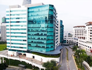
Parkway Pantai
Kuala Lumpur, Malaysia
Parkway Pantai located in Kuala Lumpur, Malaysia is accredited by JCI. Also listed below are some of the most prominent infrastructural details:
- Capacity of 335 beds
- 200+ specialist Doctors
- Critical Care Unit
- Neonatal Intensive Care Unit
- Operating Theatre
- International Patient Care Center
- Types of rooms available- Premier Suite, Supreme Suite, Deluxe Single Room, 2-bedded room, 4-bedded room, Deluxe Suite, Premier Single room, and Supreme Single room

Damansara Specialist Hospital, Petaling Jaya
Petaling Jaya, Malaysia
- KPJ Damansara Specialist Hospital offers specialised healthcare across cardiology, oncology, orthopedics, neurology, gastroenterology, nephrology, women’s health, pediatrics, ENT, and minimally invasive surgery. The hospital provides advanced diagnostics, rehabilitation, cardiac care, neurosurgery, wellness programs, and preventive healthcare supported by multidisciplinary specialists and patient-centred clinical protocols. Equipped with modern medical technologies and safety-driven systems, it focuses on evidence-based treatment, personalised care planning, and quality healthcare delivery

Prince Court Medical Centre
Kuala Lumpur, Malaysia
Prince Court is a private medical facility in the center of Kuala Lumpur that has 277 individual beds. Our dedication is to provide you with the best treatment possible from the time you arrive until you depart, healthier and happier. We are well-known for our top-notch facilities, cutting-edge technology, and friendly customer service.
Our Services to better your experience
Opinion & Option
We submit the most accurate opinion and options from one or more countries for your review
Consult Privately
Consult with a certified specialist privately on our telemedicine platform even before you decide to travel
Logistics
We handle flights, visas, transfers, and accommodation—so you can focus on your health.
Recovery
Our In-house rehabilitation service packages to better your recovery and treatment outcome
Why Choose Malaysia for Ureterolithiasis (Ureteral Stones) Treatment?
- Experienced Urology Specialists: Malaysian urology centres treat a wide range of urinary stone conditions, including ureteral stones, recurrent stone disease, obstructing stones, and complex cases.
- Advanced Diagnostic Facilities: Many hospitals offer ultrasound, CT imaging, laboratory testing, stone analysis, metabolic evaluation, and other tests to determine stone size, location, composition, and obstruction.
- Minimally Invasive Stone Treatment: Depending on the patient's condition, treatment may include ureteroscopy, laser lithotripsy, shock-wave lithotripsy, and ureteral stenting.
- Comprehensive Urological Care: Patients with recurrent or complicated stones can receive coordinated care involving urologists, radiologists, nephrologists, dietitians, and other specialists, as needed.
- International Patient Support: Major hospitals in Malaysia may provide medical-record review, specialist coordination, treatment scheduling, travel assistance, accommodation support, and follow-up arrangements for international patients.
Frequently Asked Questions
Author

Dr. Shagufta Parveen
Dr. Shagufta Parveen is a Clinical researcher and medical writer with expertise in clinical pharmacology and pharmacotherapeutics. She holds a B.Pharm and Doctor of Pharmacy (Post-Baccalaureate) degree from Teerthanker Mahaveer University, Moradabad.
During her clinical stint at BLK-Max Super Speciality Hospital and Indraprastha Apollo Hospital, she gained hands-on experience in the Clinical Pharmacology Department. Combining scientific knowledge with strong medical writing skills, Dr. Shagufta develops evidence-based healthcare content, treatment guides, and patient education resources.
Her work focuses on simplifying complex medical concepts while maintaining scientific accuracy, helping readers better understand healthcare advancements and treatment options.
In addition to her writing expertise, she is actively involved in scientific research and has contributed to peer-reviewed publications.
Her research work is accessible through the following links:
https://scholar.google.com/citations?user=lMVK1eIAAAAJ&hl=en
https://carcinogenesis.com/index.php/JOC/article/view/870
https://carcinogenesis.com/index.php/JOC/article/view/868
https://wjpsronline.com/abstract/0000000760
View More