Transitional Cell Carcinoma Treatment in India
- Top Accredited Hospitals
- Global Medical Standards
- Cutting-Edge Technology
- Proven Treatment Options
- High Success Rate
- Trusted by Millions

What is Transitional Cell Carcinoma?
Urothelial carcinoma (transitional carcinoma) begins in the urothelium, the tissue lining some areas of the urinary system. About 90% of bladder cancer cases and 7% of kidney cancer cases, including cancer of the renal pelvis and ureter, are caused by urothelial carcinoma. The symptoms of urothelial carcinoma-induced bladder and kidney cancers are comparable. Their prognoses are similar; these malignancies are easily treated when detected early but frequently return.
How does urothelial carcinoma affect my body?
Urothelial carcinoma affects the bladder and kidneys in various ways. Bladder abnormal urothelial cells can move from the lining to deeper structures, potentially travelling to distant sites such as the liver, lungs, bones, fatty tissues, and lymph nodes. Low-grade bladder cancer is not likely to metastasise, whereas high-grade disease can be lethal and might recur. Urothelial carcinoma in the kidney may progress to develop tumours in the renal pelvis or ureter, which may then extend to other organs. Both diseases, if not treated, may lead to severe implications.
What is the Importance of Timely Treatment?
Transitional cell carcinoma (TCC), also known as urothelial carcinoma, needs to be treated early to enhance prognoses and prevent the cancer from spreading. If diagnosed early, TCC is often successfully treated with less invasive methods, such as localised or transurethral resection. Postponing therapy may make treatment more difficult and reduce survival by giving the cancer time to move to other parts of the body, such as the liver, lungs, or lymph nodes, or grow deeper into the bladder. Early treatment significantly enhances the outlook for individuals with TCC, controls symptoms, and prevents recurrence.
What are the Common Symptoms of Transitional Cell Carcinoma?
Symptoms of urothelial cancer may not be present at once. Usually, the first noticeable sign is blood in the urine. You should consult a healthcare provider if you notice any symptoms, including blood in the urine.
- Chronic pain in the back.
- Fatigue.
- Unexplained weight loss.
- Dysuria is painful urination.
- Tumour or lump near your kidneys.
- Low-grade fever.
Causes and Risk Factors of Transitional Cell Carcinoma
Causes
The exact aetiology of urothelial carcinoma in the kidneys and bladder is not known to medical scientists. They have found, however, a few common risk factors:
- Cigarette smoke: Smoking cigarettes increases the risk of urothelial carcinoma-related urinary system malignancies.
- Exposuretocertainchemicals: Studies show that individuals who work with chemicals used in paint, leather, rubber, dyes, certain fabrics, and hair care products are at increased risk for urinary system malignancies linked to urothelial carcinoma.
Risk Factors
Some risks of transitional cell carcinoma are mentioned below:
- Smoking
- Exposure to chemicals
- Family history
- Genetic factors
Connect with our advisor for a PRIORITY response
Transitional Cell Carcinoma Prevention Tips
Although there is no guaranteed method to prevent Transitional Cell Carcinoma (TCC), the most important things you can do to lower your risk are to stop smoking, limit your exposure to certain chemicals at work, drink lots of water, and eat a diet high in fruits and vegetables.
Treatment options for Transitional Cell Carcinoma
Surgery is the primary treatment for localised TCC. This includes partial nephrectomy for kidney tumours and transurethral resection for bladder tumours. In certain situations, a radical cystectomy (removal of the bladder) or nephrectomy (removal of the kidney) may be necessary.
Chemotherapy: isfrequently used to eradicate any cancer cells that remain after surgery or for advanced TCC. Chemotherapy is administered intravenously or directly into the bladder for superficial bladder tumours.
Cost Start From USD 600 - USD 700Explore Options
Immunotherapy: Pembrolizumab and other immune checkpoint inhibitors are used more frequently, particularly for advanced or metastatic TCC, since they aid the immune system in recognising and combating cancer cells.
Cost Start From USD 3000 - USD 5000Explore Options
Radiation therapy is used to decrease tumours before surgery, particularly for bladder TCC, or to treat tumours in locations that are challenging for surgery to reach.
Targeted Therapy: Medications that attack cancer cells specifically based on genetic mutations or tumor-specific proteins are applied in targeted therapy for urothelial carcinoma. The medications can block the signals through which cancer cells grow and disseminate.
Cost Start From USD 8000 - USD 12000Explore Options
Urinalysis: A test to determine the colour of your urine and its components, including germs, blood, protein, and sugar.
Urine cytology: Medical professionals use a microscope to look for abnormal cells in your urine. Cancer cells may leak into your urine if you have cancer in your kidneys, bladder, or ureter.
An intravenous pyelogram (IVP) is a sequence of X-rays of the kidneys, ureter, and bladder to screen for malignancy. Medical professionals inject a contrast dye into one of the veins. To check for obstructions, physicians take X-rays as the dye passes through the kidneys, ureter, and bladder.
Ureteroscopy: To view the ureter and renal pelvis and acquire tissue samples, healthcare professionals use a narrow, tube-like device with a light and viewing lens.
Computed tomography (CT) scans provide detailed images of your internal organs by connecting a computer to an X-ray machine. Computerised tomography or computerised axial tomography are other names for this process.
Ultrasound: A process that creates echoes by reflecting high-energy sound waves off of inside organs or tissues. A sonogram is an image of bodily tissues produced by the echoes. Medical professionals may perform an abdominal ultrasound to help in the diagnosis of renal pelvic and ureter cancer.
Magnetic resonance imaging (MRI) involves creating finely detailed images of body parts, such as the pelvis, using a magnet, radio waves, and a computer. Nuclear magnetic resonance imaging (NMRI) is another name for this process.
MediRehab (chain of Rehab centres - Part of MediGence) provides comprehensive rehabilitation services designed to support Transitional cell Carcinoma patients in India. These services include:
- Physical therapy enhances strength, endurance, and mobility after chemotherapy or surgery.
- Occupational therapy: Helping the patient return to usual work and activities, especially after invasive treatment such as surgery or chemotherapy.
- Depending on the situation, your healthcare professional may prescribe medicine to help control your symptoms and support the treatment plan.
Instantly Connect with our Specialists


Hospitals for Transitional Cell Carcinoma in India

Asian Institute of Medical Sciences
Faridabad, India
Asian Institute of Medical Sciences located in Faridabad, India is accredited by NABH, NABL. Also listed below are some of the most prominent infrastructural details:
- 425 bed capacity
- Preventive and diagnostic sevices of the hospital are its strength.
- It is also well recognised for the therapeutic and rehabilitative services.
- For patients who need end process care for advanced healthcare conditions, the palliative services that the hospital provides are a boon.
- The healthcare delivery of AIMS, Delhi/NCR is focused on patient care.
- Research focused organisation
- International Patient care center is present to liason with medical travellers
- Focus on Academics, various healthcare educational programmes

BLK-Max Super Speciality Hospital
Delhi, India
BLK-Max Super Speciality Hospital, located in New Delhi, is the leading best-in-class healthcare institution in India, providing tertiary and quaternary care. 650 + beds including 162 critical care beds, 22 OTs with 1,500+ clinical professionals allied to health are at the hospital's disposal. JCI, NABH, and NABL accredited. The hospitals are a leader in transplants, cancer care, and robotic surgery education and implementation. Established in1959 by Dr. B.L. Kapur, the hospital combines state-of-the-art technology with patient-centric values including compassion, efficiency, and consistency.
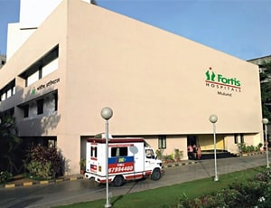
Fortis Hospital, Mulund
Mumbai, India
Fortis Hospital Mulund, located in Mulund West, Mumbai, is a premier healthcare facility providing excellence in multi-speciality healthcare for over 20 years. The global leader in advancing medical technology and clinical expertise, the hospital offers specialities including Cardiology, Neurosciences, Oncology, Transplants, and Orthopaedics. The hospital is well-known for being the largest transplant center in Maharashtra and offers advanced treatment options, including Transcatheter Aortic Valve Implantation (TAVI) and stereotactic precision radiotherapy for cancer. Fortis Mulund is equipped with a 24/7 Emergency Department and International Patient Unit to provide world-class patient-centric care.
Our Services to better your experience
Opinion & Option
We submit the most accurate opinion and options from one or more countries for your review
Consult Privately
Consult with a certified specialist privately on our telemedicine platform even before you decide to travel
Logistics
We handle flights, visas, transfers, and accommodation—so you can focus on your health.
Recovery
Our In-house rehabilitation service packages to better your recovery and treatment outcome
Why Choose India for Transitional Cell Carcinoma Treatment?
India is an excellent place for transitional cell carcinoma treatment because of its top-notch medical facilities, qualified oncologists, and best-in-class treatment facilities at reasonable prices. The nation has the latest technology-equipped state-of-the-art hospitals to facilitate effective diagnosis and treatment. Indian hospitals are also internationally accredited, offering high-quality care.
Frequently Asked Questions
Early-stage TCC kidney carcinoma may have over 90% survival, as the National Carcinoma Institute (NCI) reported. The survival rate may be 15% or below if the TCC has progressed past the bladder's inner lining or to other body parts.
The diagnosis of Transitional Cell Carcinoma, or TCC, is more prevalent among older individuals, particularly in India, who are over the age of 55. It is more prevalent among men compared to women. With age, the risk increases due to factors such as prolonged exposure to carcinogens (such as nicotine and industrial chemicals) and the additive effects of long-standing renal or bladder diseases.
Since smoking is one of the substantial risk factors for bladder, kidney, and ureter cancers, it plays a significant role in developing Transitional Cell Carcinoma (TCC) in India. Aromatic amines and various carcinogens present in tobacco smoke are absorbed into the bloodstream, filtered through the kidneys, and finally excreted in urine. These compounds can damage healthy urinary tract cells, causing mutations and the development of TCC. In India, smoking is believed to increase an individual's risk of developing TCC by as much as three times for both genders. It is one of the country's leading preventable causes of TCC.
In India, transitional cell carcinoma (TCC), especially of the bladder and ureters, can recur after treatment. TCC is a high-recurrence tumour, especially in superficial or early-stage cases, even after successful initial treatment, e.g., surgery or chemotherapy.
Regular follow-up with imaging, urine examination, and cystoscopy is mandatory to detect any recurrence at an early stage. Recurrence may be more aggressive and more challenging to manage in advanced cases. To manage the risk of recurrence, Indian patients are advised to undergo therapy and monitoring on a routine basis.
In India, radiation therapy is used to cure Transitional Cell Carcinoma (TCC) in cases where the tumour is incurable or advanced. It is also commonly used to cure bladder cancer, either alone or as a means to destroy cancer cells that remain even after surgery.
Radiation therapy can be combined with chemotherapy for better results. However, due to anatomical challenges, its use is limited to TCC, which aims to decrease symptoms, reduce tumours, and improve the overall quality of life.
Lifestyle and dietary changes can improve overall health and treatment outcomes for TCC patients in India. Some recommendations include
- Drinking plenty of water to help remove toxins and reduce the risk of recurrence.
- Consuming a diet rich in whole grains, fruits, and vegetables to enhance overall health and immunity.
- Reducing the risk of cancer by eating less processed food and red meat.
- Decreasing alcohol intake and smoking can enhance the recovery process and reduce the risk of cancer development.
- Maintaining an active lifestyle to cope with stress, build strength, and reduce fatigue.
- Enhancing mental well-being during treatment through taking part in stress-relieving activities such as yoga or meditation.

Reviewer

Dr. Mohit Agarwal
Medical Oncologist
14 Years of Experience
One of the finest Oncologist in New Delhi, India, Dr. Mohit Agarwal has worked with several world class multidisciplinary hospitals over the years. Dr. Mohit Agarwal has over 14+ years of experience in his field. The doctor treats and manages a wide range of conditions such as Stomach Cancer, Rectal Cancer, Breast Cancer, Brain Cancer View More
Last Reviewed - January 2026