Mitral Stenosis Treatment in India
- Top Accredited Hospitals
- Global Medical Standards
- Cutting-Edge Technology
- Proven Treatment Options
- High Success Rate
- Trusted by Millions

What is Mitral Stenosis?
Mitral Stenosis occurs when the opening of the mitral valve becomes narrowed, making it harder for blood to move from the left atrium into the left ventricle. Rheumatic heart disease is the most common cause worldwide, although mitral stenosis can also result from valve calcification, congenital abnormalities, or other less common conditions. Severe disease can lead to pulmonary hypertension, atrial fibrillation, heart failure, and other complications.
What is the Importance of Timely Treatment?
Timely treatment can help relieve breathlessness and other symptoms, reduce pressure on the heart and lungs, manage complications such as atrial fibrillation, and prevent worsening heart failure. Regular assessment also helps determine whether monitoring, medicines, percutaneous mitral balloon commissurotomy, or surgery may be appropriate based on valve anatomy and disease severity.
What are the Common Symptoms of Mitral Stenosis?
- Shortness of Breath: Breathlessness may occur during physical activity and can become more noticeable as the valve narrowing progresses.
- Fatigue: Reduced blood flow into the left ventricle may contribute to tiredness and reduced physical endurance.
- Palpitations: An irregular or rapid heartbeat may occur, particularly when atrial fibrillation develops.
- Reduced Exercise Tolerance: Routine activities may become increasingly difficult because of breathlessness or fatigue.
- Swelling: Fluid retention may cause swelling of the ankles, feet, or lower legs, particularly in more advanced disease.
- Dizziness or Chest Discomfort: Some patients may experience lightheadedness or chest discomfort, especially during exertion.
Causes and Risk Factors of Mitral Stenosis
Causes
- Rheumatic Heart Disease: Previous rheumatic fever can cause progressive scarring and narrowing of the mitral valve and remains the most common cause worldwide.
- Mitral Valve Calcification: Calcium deposits can progressively restrict valve movement, particularly in older adults.
- Congenital Mitral Stenosis: Some people are born with mitral valve abnormalities that cause narrowing.
- Autoimmune or Inflammatory Conditions: Conditions such as lupus can rarely contribute to mitral valve disease.
- Other Structural Valve Changes: Less common abnormalities may also interfere with normal mitral valve opening.
Risk Factors
- Previous rheumatic fever or rheumatic heart disease
- Increasing age
- History of valvular heart disease
- Congenital heart abnormalities
- Conditions associated with valve calcification
- Certain autoimmune or inflammatory diseases
- Previous heart-valve procedures
Latest Research and Technologies in the Treatment of Mitral Stenosis in India
- Current advances in mitral stenosis care include multimodality cardiac imaging, three-dimensional echocardiography, transesophageal echocardiography, cardiac CT, and advanced catheter-based assessment to define valve anatomy and treatment suitability.
Treatment options for Mitral Stenosis
Percutaneous Mitral Balloon Commissurotomy (PMBC): Also known as Percutaneous Mitral Valvuloplasty (PMV), this is a catheter-based, first-line mechanical treatment to split open fused leaflets in patients with favourable rheumatic anatomy (measured via a low Wilkins score).
Mitral Valve Replacement (MVR) : The standard open-surgical option using either a mechanical or bioprosthetic valve, reserved for patients with severe calcification, distorted subvalvular apparatus, or concurrent severe mitral regurgitation.
Cost Start From 4000Explore Options
Transcatheter Mitral Valve Replacement (TMVR): This specialised procedure (such as valve-in-mitral-annular-calcification, or ViMAC) is an emerging option for elderly, high-risk patients with degenerative disease who cannot undergo open surgery.
- Clinical assessment:
- Medical History: Assessment of breathlessness, fatigue, exercise tolerance, palpitations, previous rheumatic fever, heart disease, and medications.
- Physical Examination: Evaluation of heart sounds, heart rhythm, blood pressure, oxygen saturation, signs of fluid retention, and other cardiovascular findings.
- Electrocardiogram (ECG): May identify atrial fibrillation, heart-rate abnormalities, or other cardiac changes.
- Blood Tests: Blood tests may assess kidney function, electrolytes, blood counts, and other factors relevant to treatment.
- Exercise Testing: Exercise or stress testing may be considered when symptoms and resting valve measurements do not clearly correspond.
- Imaging Tests:
- Transthoracic Echocardiography (TTE): The standard initial imaging test used to assess valve structure, valve area, blood-flow gradients, heart chambers, and pulmonary pressures.
- Transesophageal Echocardiography (TEE): Provides detailed images of the mitral valve and left atrium and may be used before percutaneous commissurotomy to assess for blood clots and valve anatomy.
- Three-Dimensional Echocardiography: Provides detailed assessment of valve morphology and can support procedural planning in selected patients.
- Cardiac CT: May provide additional anatomical information, particularly when valve or mitral annular calcification needs further assessment.
- Cardiac MRI: Provides detailed information about heart structure and function in selected patients.
- Chest X-ray: May show enlarged cardiac chambers or changes associated with pulmonary congestion.
- Cardiac Catheterisation: Can measure pressures within the heart and pulmonary circulation when non-invasive testing does not provide sufficient information or before selected interventions.
MediRehab (a chain of Rehab centres, part of MediGence) provides comprehensive rehabilitation services designed to support Mitral Stenosis patients in India. These services include:
- Cardiac Rehabilitation: Structured rehabilitation may help improve exercise capacity and support recovery after valve procedures.
- Gradual Physical Activity: Increase activity according to symptoms, valve severity, heart function, and medical advice.
- Breathing and Functional Training: Selected patients may benefit from supervised exercises to improve functional capacity.
- Long-Term Cardiac Follow-Up: Regular cardiology and valve-clinic assessments help monitor valve function, heart rhythm, symptoms, and treatment response.
- Medicines for Mitral Stenosis may include diuretics, heart-rate controlling medicines, medicines for atrial fibrillation, and anticoagulants when indicated. These treatments can reduce symptoms and manage complications but do not reverse severe mechanical narrowing of the mitral valve. In rheumatic mitral stenosis with atrial fibrillation, anticoagulation decisions require particular consideration because thromboembolic risk is increased.
Instantly Connect with our Specialists


Hospitals for Mitral Stenosis in India

Yatharth hospital
Delhi, India
- The hospital features over 400 beds with advanced infrastructure for multi-speciality treatment.
- Much of the facility is dedicated to ICU units with the latest life-saving equipment.
- Advanced medical imaging systems include a 64-slice cardiac CT scanner, a dual-head gamma camera, a 16-slice PET-CT scanner, and a fan-beam bone density scanner (BMD).
- The radiology setup operates through a fully integrated RIS-HIS system for smooth diagnostics and reporting.
- The hospital offers high-resolution colour Doppler ultrasound systems for precise vascular and soft tissue imaging.
- The cardiology wing is equipped with a Philips FD20/10 cath lab, enhanced by modern stent-boost imaging technology.
- Cutting-edge tools, such as optical coherence tomography (OCT), intravascular ultrasound (IVUS), and fractional flow reserve (FFR), support advanced heart diagnostics.
- Facilities include rotablator technology for hardened arteries and an Ensite Velocity cardiac mapping system for complex arrhythmias.
- A fully functional hybrid endovascular suite supports combined surgical and interventional heart procedures.
- The ICU and NICU areas feature high-frequency ventilators, central pressure monitoring, and advanced bedside diagnostic tools.
- Life-support systems include intra-aortic balloon pumps, invasive blood pressure monitoring, and bedside percutaneous tracheostomy.
- Patient safety and comfort are enhanced with temperature-controlled blankets, bedside echocardiography, and mobile X-ray systems.
- Operation theatres are fitted with navigation-assisted systems for knee replacement and MEP monitoring for spine surgeries.
- Fiberoptic bronchoscopes, PCA pumps for pain control, and SSEP monitoring during neurosurgeries enhance surgical precision.

Global Health City
Chennai, India
The Hospital has world-class infrastructure with a capacity of over 1000 beds and much more-
- 265 Licensed beds
- 13 Operation Theatres
- 24*7 Cath Lab
- 24*7 available Blood Bank
- 24*7 Emergency Department
- 24*7 Open Pharmacy
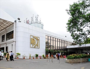
Apollo Hospitals
Hyderabad, India
Apollo Hospitals located in Hyderabad, India is accredited by JCI, NABH. Also listed below are some of the most prominent infrastructural details:
- A premier multi-specialty hospital with 477-bed capacity
- More than 50 specialties, super-specialties
- 12 Centers of Excellence
- The Institutes for Heart Diseases, Neurosciences, Cancer, Emergency, Orthopaedics, Renal Diseases, and Transplants
- Centers of Excellence are known for patient care, training and research
- Doctors with years of truly global experience in healthcare delivery
Our Services to better your experience
Opinion & Option
We submit the most accurate opinion and options from one or more countries for your review
Consult Privately
Consult with a certified specialist privately on our telemedicine platform even before you decide to travel
Logistics
We handle flights, visas, transfers, and accommodation—so you can focus on your health.
Recovery
Our In-house rehabilitation service packages to better your recovery and treatment outcome
Why Choose India for Mitral Stenosis Treatment?
- Experienced Cardiac Specialists: Access to cardiologists, interventional cardiologists, and cardiac surgeons experienced in managing valvular heart disease.
- Advanced Valve Diagnostics: Major cardiac centres may offer advanced echocardiography, 3D imaging, cardiac CT, cardiac MRI, and catheter-based assessment.
- Percutaneous Valve Procedures: Eligible patients may have access to percutaneous mitral balloon commissurotomy and other catheter-based interventions.
- Cardiac Surgery: Specialised centres can provide mitral valve repair, commissurotomy, and valve replacement when required.
- Multidisciplinary Heart Teams: Cardiologists, cardiac surgeons, imaging specialists, anaesthesiologists, and rehabilitation teams can collaborate on complex cases.
- Personalised Treatment Planning: Treatment selection can consider valve anatomy, symptoms, pulmonary pressures, heart rhythm, age, associated conditions, and procedural risk.
- Comprehensive Follow-Up: Long-term monitoring can help manage valve function, rhythm abnormalities, anticoagulation, and other complications.
- International Patient Support: Many major Indian hospitals assist international patients with appointments, treatment coordination, accommodation, and follow-up.