Arteriovenous Malformation (AVM) Treatment in India
- Top Accredited Hospitals
- Global Medical Standards
- Cutting-Edge Technology
- Proven Treatment Options
- High Success Rate
- Trusted by Millions

What is an Arteriovenous Malformation (AVM)?
An arteriovenous malformation (AVM) is an abnormal connection or tangled network of arteries and veins in which blood flows directly from arteries into veins without passing normally through capillaries. AVMs can occur in different parts of the body but are commonly associated with the brain and spinal cord. This abnormal blood flow can stress the affected vessels and, in some cases, lead to bleeding or damage to surrounding tissues.
What is the Importance of Timely Treatment?
Timely evaluation is important because some AVMs, particularly those in the brain or spinal cord, can rupture and cause bleeding, neurological damage, seizures or stroke-like complications. Treatment decisions are individualised based on the AVM's location, size, symptoms, bleeding history, and the risks of intervention.
What are the Common Symptoms of an Arteriovenous Malformation (AVM)?
- Headache
- Seizures
- Weakness or numbness
- Problems with balance or coordination
- Vision disturbances
- Speech or language difficulties
- Dizziness
- Pain, particularly with some spinal AVMs
- Pulsatile tinnitus in some brain AVMs
- Confusion or loss of consciousness
- Neurological symptoms associated with bleeding
Causes and Risk Factors of Arteriovenous Malformation (AVM)
Causes
- The exact cause of most AVMs is not known. Many are believed to develop during vascular formation before birth, although symptoms may not appear until childhood or adulthood.
- Certain inherited vascular conditions, including hereditary hemorrhagic telangiectasia and capillary malformation-AVM syndromes, can be associated with AVMs.
Risk Factors
- Congenital or developmental vascular abnormalities
- Family or genetic history of certain vascular disorders
- Hereditary hemorrhagic telangiectasia (HHT)
- Capillary malformation-AVM syndromes
- Presence of a previously identified AVM
- Certain inherited vascular conditions
Latest Research and Technologies in the Treatment of Arteriovenous Malformation (AVM) in India
- Advances in AVM care include high-resolution MRI and angiographic imaging, advanced endovascular embolisation techniques, microsurgical approaches and stereotactic radiosurgery. For selected brain AVMs, 3D imaging and treatment-planning technologies can help clinicians understand the relationship between the AVM and surrounding brain structures. Embolisation may be used alone or combined with surgery or radiosurgery, while stereotactic radiosurgery can gradually close abnormal vessels over time.
Treatment options for Arteriovenous Malformation (AVM)
Endovascular Embolisation : A catheter is guided into vessels supplying the AVM, where an embolizing material is delivered to reduce or block abnormal blood flow. It may be used alone or before surgery or radiosurgery.
Cost Start From 12500Explore Options
Microsurgical Resection: Selected AVMs, particularly small ones in surgically accessible areas, may be removed through microsurgery. The aim is complete removal while preserving surrounding healthy tissue.
Gamma Knife Radiosurgery (GKRS) : A premier, non-invasive treatment option for Cerebral Arteriovenous Malformations (AVMs). Rather than utilising a physical blade, it targets the precise tangled web of abnormal vessels (the nidus) using hundreds of highly focused beams of gamma radiation.
Cost Start From 5000Explore Options
Combined Treatment: Some complex AVMs require a staged or combined approach, such as embolisation followed by surgery or radiosurgery. The treatment plan depends on the vascular anatomy and individual risk profile.
Observation and Regular Monitoring: Some AVMs may be monitored with periodic imaging when immediate intervention is not considered appropriate. Follow-up helps assess changes and guide future management.
- Clinical assessment:
- Detailed medical and family history, neurological or physical examination, and assessment of symptoms such as headaches, seizures, weakness, sensory changes, vision problems or previous bleeding.
- Doctors may also evaluate for associated inherited vascular conditions when clinically indicated.
- Imaging Tests:
- MRI/MRA: Provides detailed images of the AVM and surrounding tissues and helps assess its location and characteristics.
- CT/CT Angiography: May identify bleeding and provide information about the blood vessels and surrounding structures.
- Cerebral Angiography: Provides detailed visualization of feeding arteries, the AVM nidus and draining veins and is important for treatment planning in brain AVMs.
- Spinal Angiography: May be used when a spinal AVM is suspected to define its vascular anatomy.
- Ultrasound/Doppler: May be useful for selected AVMs in superficial or accessible parts of the body.
- Functional or specialized imaging: Additional imaging may be used to assess the AVM's relationship with important brain or spinal structure
MediRehab (a chain of Rehab centres, part of MediGence) provides comprehensive rehabilitation services designed to support Arteriovenous Malformation (AVM) patients in India. These services include:
- Physiotherapy: Helps improve strength, balance, coordination and mobility when neurological deficits affect movement.
- Occupational Therapy: Helps patients regain independence with daily activities and adapt to physical or neurological limitations.
- Speech and Language Therapy: May be recommended for patients who develop speech, language or swallowing difficulties following neurological complications.
- Neurorehabilitation: A structured rehabilitation programme can help address cognitive, motor and functional problems after AVM-related bleeding or treatment.
- Long-Term Follow-Up: Regular neurological assessment and follow-up imaging may be required to monitor recovery and confirm treatment response.
- Medicines usually do not eliminate the AVM itself but may control associated symptoms or complications. Antiseizure medicines may be prescribed for patients experiencing seizures, while pain-relieving medicines may help manage headaches or pain associated with certain AVMs. Following AVM-related bleeding or a neurological complication, additional medicines may be prescribed according to the patient's condition. The treating specialist should individualise medication decisions.
Instantly Connect with our Specialists


Hospitals for Arteriovenous Malformation (AVM) in India

Apollo Hospital
Chennai, India
Apollo Hospital located in Chennai, India is accredited by JCI, NABH. Also listed below are some of the most prominent infrastructural details:
- Committed Centers of Excellence involving many major specialties, super specialties
- Assistance in trip planning and execution
- Insurance related assistance
- Visa facilitation
- International patient representatives for complete end to end travel and transfer processing for medical travellers
- Language translators availability
- Robust safety and infection protocols
- Personalised, Visa and Premium Health Checks available
- Health Library and accessing health records online
- Wide variety of procedures accomplished including complex and critical procedures
- Technologically advanced systems and procedures in place
- Research and academics base of healthcare delivery

Indraprastha Apollo Hospital
Delhi, India
Indraprastha Apollo Hospital is known for delivering treatment to over 200,000 patients every year; 10,000 of which are generally medical tourists. The efficient team of doctors has the record of 99.6 percent success rate. Indraprastha Apollo Hospital deals in treatment of over 50 specialities.
Let’s see some of the features of the infrastructure:
- Spread over 15 acres, the hospital has 710 beds.
- 6 beds dedicated to bone marrow transplant units with very strict infection control practices.
- 64-Slice Scan coupled with data acquisition which provides highest temporal resolution
- One of the hospitals to be using Da Vinci Robotics Surgery System
- In South Asia, the Spect-CT and Pet-CT got their first installation at Indraprastha Apollo Hospital in India
- Technologies such as PET- MR, BrainLab Navigation System, PET-CT, Portable CT Scanner, Tilting MRI, NovalisTx, Cobalt based HDR Brachytherapy, Hyperbaric Chamber, DSA Lab, Fibroscan, 3 Tesla MRI, Endosonography, 128 Slice CT scanner are all installed at the hospital.
- The Cancer Institute at Indraprastha Apollo is equipped with a highly advanced Radiation Oncology Center with ClinaciX, NovalisTx, and HDR-Brachytherapy.
- It is equipped with the largest Sleep Lab in Asia, & has one of the largest Dialysis Units in India.
- A large number of ICU beds than any other Private Hospitals in India.
- WIFI is accessible across the whole campus.
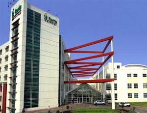
Fortis Hospital
Noida, India
Fortis Healthcare Limited is one of India’s prominent providers of integrated healthcare services, offering comprehensive medical care across multiple specialities. The company’s operations span hospitals, diagnostic centres, and day-care speciality facilities, ensuring a wide range of healthcare solutions under one umbrella. Fortis has established a strong presence across the country, managing 33 healthcare facilities, which include joint ventures and operational & management (O&M) setups, across 11 states.
The company’s hospital network comprises more than 5,700 operational beds, including O&M beds, catering to patients with varied healthcare needs. In addition to inpatient services, Fortis operates around 400 diagnostic laboratories, providing reliable and timely diagnostic support for accurate medical care. This extensive network enables Fortis to deliver high-quality healthcare services consistently and efficiently, from routine diagnostics to specialised treatments and complex procedures.
Our Services to better your experience
Opinion & Option
We submit the most accurate opinion and options from one or more countries for your review
Consult Privately
Consult with a certified specialist privately on our telemedicine platform even before you decide to travel
Logistics
We handle flights, visas, transfers, and accommodation—so you can focus on your health.
Recovery
Our In-house rehabilitation service packages to better your recovery and treatment outcome
Why Choose India for Arteriovenous Malformation (AVM) Treatment?
- Multidisciplinary AVM Care: Complex cases can be evaluated by neurosurgeons, neurologists, interventional neuroradiologists, radiation specialists and rehabilitation professionals.
- Advanced Vascular Imaging: Major hospitals may provide MRI/MRA, CT angiography and catheter-based cerebral angiography for detailed AVM assessment.
- Endovascular Treatment Options: Interventional neuroradiology centres may offer catheter-based embolisation for appropriately selected AVMs.
- Advanced Neurosurgical and Radiosurgical Care: Selected patients may have access to microsurgical resection and stereotactic radiosurgery, depending on AVM characteristics.
- Comprehensive Rehabilitation: Patients with neurological complications can receive physiotherapy, occupational therapy, speech therapy and neurorehabilitation as required.